
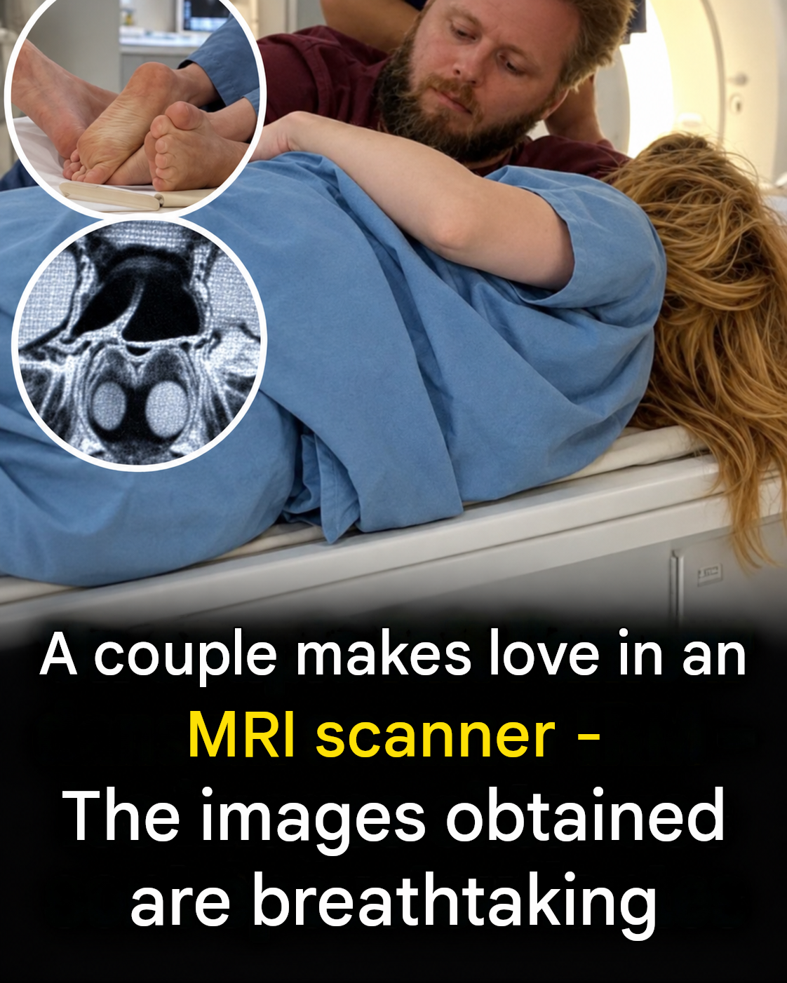
Medical imaging technology has revolutionized modern healthcare by allowing physicians to peer inside the human body with astonishing clarity. From X-rays and ultrasounds to computed tomography (CT) scans and magnetic resonance imaging (MRI), these tools help diagnose conditions, monitor treatments, and advance medical science. Occasionally, researchers and scientists utilize these powerful imaging systems in unique, unconventional studies to explore human anatomy and physiological functions in real time.
When fascinating stories or scientific studies regarding MRI scans circulate online, they instantly capture public curiosity and spark widespread discussion. Understanding how MRI technology works, how medical imaging contributes to science, and how diagnostic tools are utilized safely is both educational and intriguing.
In this comprehensive guide, we will explore the science behind MRI scanners, examine how advanced medical imaging works, discuss safety protocols in radiology, and provide practical tips for preparing for diagnostic medical scans.
The Science and Power of MRI Technology
Magnetic resonance imaging (MRI) is a sophisticated medical diagnostic tool that uses powerful magnets and radio waves to generate detailed images of the organs and tissues inside the body.
1. How Magnetic Fields Create Images
Unlike X-rays or CT scans, which use ionizing radiation, MRI machines utilize a strong magnetic field and radio frequency pulses to align hydrogen atoms in the body’s water molecules. When the pulses are turned off, the atoms emit signals that specialized computers translate into high-resolution cross-sectional images.
2. Real-Time Physiological Studies
Throughout history, medical researchers have occasionally used MRI scanners to study dynamic physiological processes in real time—ranging from joint movements and swallowing mechanics to cardiovascular function and neurological activity.
+------------------------------------+---------------------------------------+
| MRI Technology Feature             | Medical & Scientific Application      |
+------------------------------------+---------------------------------------+
| Strong Magnetic Fields             | Aligns hydrogen atoms for clear scans |
| Radio Frequency Pulses             | Generates detailed cross-sections     |
| Real-Time Imaging                  | Studies dynamic physiological actions |
| Zero Ionizing Radiation            | Ensures safety during frequent scans  |
+------------------------------------+---------------------------------------+
Safety Protocols in Medical Imaging
Because MRI machines generate immensely powerful magnetic fields, strict safety guidelines must be followed in clinical environments.
1. Eliminating Metal Objects
Patients and staff must remove all metallic items—such as jewelry, watches, keys, and clothing with metal threads—before entering the scan room, as the magnet can attract metallic objects with dangerous force.
2. Screening for Medical Implants
Individuals with certain pacemakers, metallic surgical clips, or implanted medical devices must undergo rigorous screening, as the magnetic field can interfere with or displace these items.
Practical Tips for Your Next MRI Scan
If you are scheduled for an upcoming diagnostic MRI scan, these practical tips can help ensure a smooth and comfortable experience:
Communicate Any Anxiety
If you experience claustrophobia or anxiety inside enclosed spaces, inform your doctor beforehand. They can prescribe mild sedatives or offer open-MRI alternatives if available.
Wear Comfortable, Metal-Free Clothing
Opt for loose, comfortable clothing free of zippers, snaps, or metallic embellishments, or change into the provided hospital gown.
Follow Technologist Instructions
Remain as still as possible during the scanning sequences to ensure the imaging computer captures crisp, clear diagnostic pictures.
Fascinating Facts About Medical Imaging
- The Strength of MRI Magnets: Standard clinical MRI magnets are measured in teslas (T). A typical 1.5T or 3T MRI machine is roughly 30,000 to 60,000 times stronger than the Earth’s magnetic field.
- The Noisy Environment: MRI scanners produce loud tapping, clicking, and humming noises as the internal magnetic coils switch rapidly on and off, which is why patients wear earplugs or headphones.
- Inventing the Technology: The foundational physics behind magnetic resonance imaging earned the Nobel Prize in Physiology or Medicine in 2003 for its monumental impact on healthcare.
Frequently Asked Questions (FAQ)
How does an MRI scanner work without radiation?
MRI scanners use powerful magnets and radio waves rather than ionizing radiation (such as X-rays) to map the distribution of water molecules inside the human body.
Why is it important to remove all metal before an MRI?
The magnetic pull of an MRI machine is extremely powerful and can turn metallic objects into dangerous projectiles or heat up conductive metals, causing injury.
Can an MRI scan be used to study internal organ movement?
Yes, modern fast-imaging MRI sequences allow physicians and researchers to observe dynamic movements, blood flow, and organ functions in real time.
What should I expect during an MRI procedure?
You will lie on a sliding table that moves inside a cylindrical tube while the machine produces loud tapping noises. The procedure is entirely painless and typically lasts between 20 to 60 minutes.
Are there alternatives for patients with severe claustrophobia?
Yes, wide-bore MRI machines and open-MRI scanners provide more space and comfort for patients who struggle with enclosed spaces.
Conclusion
Advanced medical imaging technologies like MRI scanners continue to push the boundaries of science, offering breathtaking insights into human anatomy and physiological functions. While viral internet stories often highlight unusual experiments, the true power of MRI lies in its ability to diagnose medical conditions and save lives every day.
Combine curiosity about medical technology with proper healthcare awareness to appreciate the incredible innovations shaping modern medicine!
