
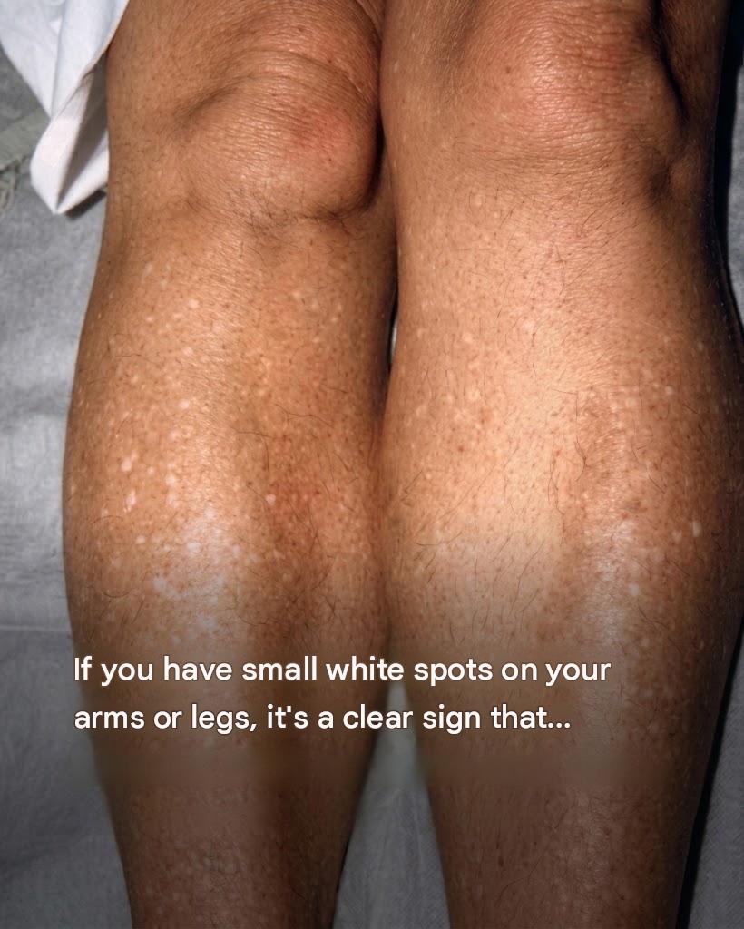
Discovering small, porcelain-white spots on sun-exposed skin—particularly on the shins, forearms, shoulders, and chest—is extremely common, especially as people age. While these flat, smooth macules can trigger concern about conditions like vitiligo or fungal infections, they are most frequently a harmless dermatological condition known as Idiopathic Guttate Hypomelanosis (IGH).
Social media posts often sensationalize these spots, falsely linking them to severe systemic illnesses or nutrient deficiencies. In reality, IGH represents a localized reduction in melanin (pigment) production, primarily resulting from cumulative, long-term sun exposure and natural cellular aging.
This comprehensive guide breaks down the causes of idiopathic guttate hypomelanosis, compares it to other skin hypopigmentation conditions, details dermatological treatment options, and provides key insights into health insurance, preventive care, and financial wellness.
1. What is Idiopathic Guttate Hypomelanosis (IGH)?
Understanding the breakdown of the medical term helps demystify the condition:
- Idiopathic: Unknown or complex underlying cause.
- Guttate: Teardrop-shaped or small, distinct spots (typically 1 to 5 millimeters in diameter).
- Hypomelanosis: Localized loss of melanin (skin pigment).
Clinical Characteristics
IGH spots typically appear as flat, smooth, porcelain-white or off-white macules. They occur most frequently on the anterior shins (front of the lower legs), forearms, upper arms, and neck—areas that receive high cumulative sun exposure throughout a person’s life. They are asymptomatic, meaning they do not itch, burn, or cause pain, and they do not transform into skin cancer.
2. Primary Causes and Biological Mechanisms
While the exact trigger remains subject to research, several key factors contribute to the localized loss of pigment cells in IGH:
1. Cumulative Sun Exposure (Photoaging)
Ultraviolet (UV) radiation from sunlight or tanning beds causes cumulative damage to melanocytes—the specialized cells responsible for producing melanin. Over time, chronic UV exposure leads to localized melanocyte destruction or a decrease in their ability to synthesize pigment.
2. Natural Cellular Aging
As the body ages, the total number of functional melanocytes naturally decreases by about 10% to 20% per decade. This gradual reduction, combined with sun exposure, causes small areas of skin to lose their pigment entirely.
3. Genetic Predisposition
Genetics plays a noticeable role; individuals with a family history of IGH are significantly more likely to develop white spots as they enter middle age.
3. Differential Diagnosis: IGH vs. Other Skin Conditions
Not all white spots on the skin are IGH. A dermatologist can differentiate IGH from other common hypopigmentation conditions:
1. Vitiligo
- Characteristics: Vitiligo is an autoimmune condition where the immune system attacks melanocytes.
- Differences: Unlike the tiny, discrete 1–5 mm dots of IGH, vitiligo causes larger, expanding chalk-white patches that often appear symmetrically around joints, eyes, mouth, or hands.
2. Tinea Versicolor (Pityriasis Versicolor)
- Characteristics: A common superficial fungal infection caused by an overgrowth of natural skin yeast (Malassezia).
- Differences: Tinea versicolor spots are often slightly scaly, mildly itchy, and merge together into larger patches on the trunk, back, or chest. It responds well to topical antifungal treatments.
3. Pityriasis Alba
- Characteristics: Frequently seen in children and young adults, especially those with eczema or atopic dermatitis.
- Differences: Appears as dry, slightly raised, poorly defined pale patches primarily on the face and upper arms, which typically resolve over time with moisturization.
4. Treatment Options and Dermatological Management
Because IGH is entirely benign, medical treatment is cosmetically optional. While reversing lost pigment completely is challenging, several dermatological options can diminish their appearance:
Dermatological Procedures
- Topical Retinoids: Prescription tretinoin or retinol creams stimulate cell turnover and may improve skin texture and mild repigmentation over several months.
- Cryotherapy: Precise, light liquid nitrogen application can occasionally stimulate melanocyte migration to the area, though cautious application is necessary to avoid scarring.
- Microdermabrasion & Chemical Peels: Superficial resurfacing treatments remove dead surface cells and encourage refreshed epidermal growth.
- Laser & Light Therapy: Fractional lasers and excimer lasers have shown success in stimulating pigment production in select cases.
Everyday Skincare & Prevention
- Daily Broad-Spectrum Sunscreen: Applying broad-spectrum SPF 30+ daily prevents new spots from forming and stops existing spots from contrasting further against surrounding tanned skin.
- Avoiding Tanning Beds: Tanning accelerates melanocyte damage and worsens the appearance of white spots.
5. Preventive Care, Health Insurance, and Financial Wellness
Navigating dermatological health thoughtfully involves maximizing healthcare benefits, routine skin screenings, and smart financial management.
Leveraging Health Coverage for Skin Health
- Annual Dermatological Screenings: Because IGH indicates significant cumulative sun exposure, individuals with numerous white spots have a higher baseline risk for other sun-related conditions, including skin cancers (such as basal cell carcinoma, squamous cell carcinoma, or melanoma). Annual full-body skin checkups with a board-certified dermatologist are routinely covered under standard health insurance plans.
- Cosmetic vs. Medical Coverage: Treatments performed purely for cosmetic appearance (such as laser therapy for IGH) are typically out-of-pocket expenses. However, consultations and diagnostic evaluations to rule out conditions like vitiligo or fungal infections are covered under standard medical benefits.
- HSA/FSA Utilization: Health Savings Accounts (HSAs) and Flexible Spending Accounts (FSAs) can often be used to purchase eligible sun protection products, prescription topical treatments, and covered clinical co-pays.
6. Fascinating Facts About Skin Pigmentation and Sun Exposure
- Melanin Shield: Melanin acts as a natural biological umbrella, absorbing harmful UV rays and protecting epidermal DNA from nuclear radiation damage.
- Epidermal Renewal: Your skin sheds roughly 30,000 to 40,000 dead skin cells every single minute, replacing its entire outer layer (epidermis) every 28 to 40 days.
- Prevalence of IGH: Clinical studies estimate that over 80% of adults above the age of 70 have at least a few small IGH spots on their lower legs or forearms.
7. Frequently Asked Questions (FAQ)
Are small white spots on arms and legs dangerous?
No. Idiopathic guttate hypomelanosis (IGH) is completely benign, non-contagious, and does not progress into skin cancer.
Can calcium or vitamin deficiencies cause white spots on the skin?
No. A common myth suggests white spots indicate a calcium or vitamin deficiency, but IGH is caused by melanocyte loss from sun exposure and aging, not dietary deficiencies.
Will sunscreen make IGH spots go away?
Sunscreen will not reverse existing spots, but it stops surrounding skin from tanning (which minimizes contrast) and prevents new melanocyte damage and new spots from forming.
Can IGH spots be permanently cured?
There is no guaranteed cure to restore 100% of lost pigment in every spot, but targeted treatments like topical retinoids or laser therapy can reduce their visibility.
Conclusion: Embrace Healthy Sun Habits and Skin Protection
Small white spots on the arms and legs are a natural reflection of time spent in the sun throughout life. Known as idiopathic guttate hypomelanosis, these spots are harmless indicators of sun exposure rather than serious medical issues.
By practicing daily sun protection, staying informed about skin health, and scheduling regular preventive checkups with a dermatologist, you can maintain healthy, resilient skin for years to come.
